Sectoral Heterochromia: How Rare It Is & What Causes It
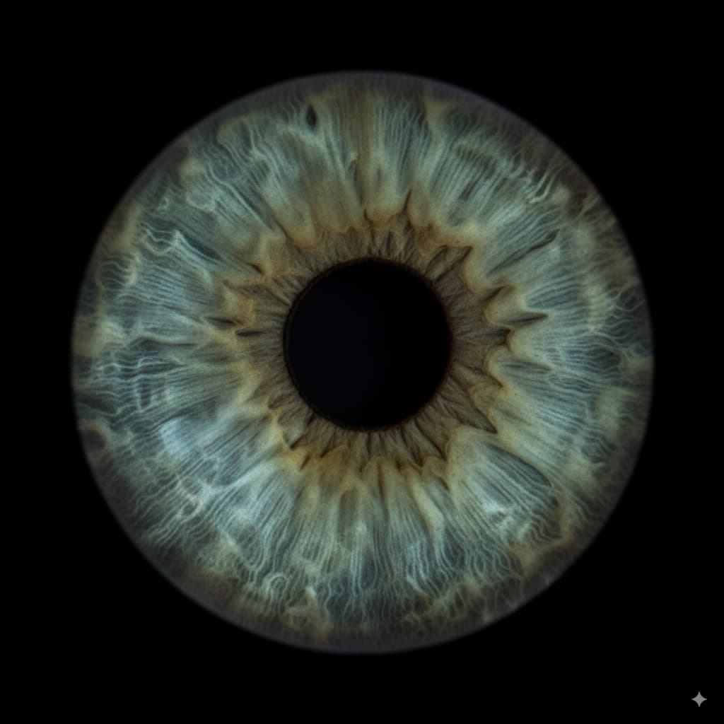
Sectoral heterochromia is a block of one color sitting inside an iris of another color. A wedge of brown in a blue eye. A slice of green in a hazel one. Most people meet it by accident, in a close-up photo of their own face, and the first thing they ask is whether it is rare.
This guide gives the honest answer to that. It also covers what causes the patch, how to tell it from an iris freckle, and the one situation that is worth an eye exam.
What Is Sectoral Heterochromia?
Heterochromia means having more than one color in the eyes. Sectoral heterochromia is the type where one iris carries a distinct block of a second color. Doctors also call it partial heterochromia, or iris sector heterochromia.
The shape is the giveaway. A sector is a slice, like a piece cut from a pie. It usually starts at the pupil and runs outward to the rim. Some are a thin sliver. Some cover a quarter of the iris. A few cover close to half.
It can show up in one eye or in both. When it shows in both, the patches are often in different places and different sizes. That is normal for this type.
The cause of the color itself is simple. Melanin is the pigment that sets eye color. More melanin reads as brown, less reads as green, blue or gray. In sectoral heterochromia, one wedge of the iris ended up with a different amount of melanin than the rest.
How Rare Is Sectoral Heterochromia?
Here is the honest answer: nobody knows, and any page that gives you a precise percentage made it up.
We went looking for a real number. A search of PubMed, the main index of medical research, for a study of how common sectoral heterochromia is in people returns nothing useful. There is no population survey. There is no clinic audit. There is no national figure.
What the literature holds instead is case reports. Search the journals for papers with sector heterochromia in the title and you get a handful, each describing one patient or one family. A case report tells you that something happened once. It cannot tell you how often it happens.
The closest thing to a real count is a study of dogs. In 1992, a team tested 900 Dalmatians for deafness and recorded heterochromia at the same time. It is a careful piece of work. It is also about dogs, and about heterochromia in general rather than the sectoral type.
So why has nobody counted it in people? Three reasons stand out.
- There is no agreed cut-off. Nobody has set a rule for how big or how different a patch has to be before it counts. Without a rule, two researchers would not count the same eyes.
- Most patches are small. A thin wedge gets called a freckle, or a streak, or nothing at all. It never reaches a doctor's note.
- You have to look closely to see it. Big health surveys record things like vision and eye pressure. They do not photograph each iris in daylight and grade its color zones.
Our main heterochromia guide quotes the figure people cite most often for heterochromia of all types: fewer than 1% of people, or roughly 6 in 1,000. That number is itself an estimate, and we label it as one. We are not going to split it into a sectoral share. Nobody has measured how that 1% divides between the three types, so any split would be invented.
What we can say, and how sure we are
One thing can be said with some confidence, and we flag it as a judgement rather than a measurement. Complete heterochromia, where one whole eye differs from the other, is the type that gets noticed and written up. Central heterochromia, a ring around the pupil, is the type most often missed or filed as hazel. Sectoral sits between them. That ordering is the common clinical impression, and it matches how often each type gets reported. It is not a counted result, so treat it as a rough sense of scale rather than a fact.
Sectoral, Central or Complete: Which One Do You Have?
| Type | What you see | Which eyes |
|---|---|---|
| Sectoral | A wedge or patch of a second color inside one iris, usually running out from the pupil | One eye, sometimes both, and often in different spots |
| Central | A ring of one color around the pupil, a different color toward the rim | Almost always both eyes, matching |
| Complete | One whole iris is a different color from the other | Both eyes, each a single color |
The quick test is the edge. A sector has a border you can trace, and the color on each side of that border is different. A central ring is a circle, not a wedge. Complete heterochromia needs no test at all.
If your two eyes look the same but each has a bright ring near the pupil, read our guide to central heterochromia instead. If the colors blur together across the whole iris with no border, you are probably looking at hazel eyes.
Sectoral Heterochromia or an Eye Freckle?
This is the mix-up we see most, and the two are genuinely easy to confuse. Both put a patch of a different color on the iris. They are not the same thing.
An iris freckle, or a nevus, is a spot of extra pigment sitting on or in the iris. It is almost always darker than the iris around it, and it is usually small, rounded and set somewhere in the middle of the color ring.
Sectoral heterochromia is a section of the iris itself that formed with a different amount of pigment. It tends to be wedge shaped. It often reaches from the pupil to the outer rim. And it can be lighter than the rest of the eye as easily as darker.
| What to look at | Iris freckle or nevus | Sectoral heterochromia |
|---|---|---|
| Shape | A blob or a dot | A wedge or a slice |
| Direction | Sits wherever it sits | Usually points outward from the pupil |
| Which way the color goes | Nearly always darker than the iris | Can be lighter or darker |
| Size | Small | Often big enough to change how the whole eye reads |
Neither of these is something you should settle by yourself. Our guide to eye freckles covers what they are and when a spot needs a look. If you are unsure which one you have, that is a question for an eye exam, not a web page.
What Causes Sectoral Heterochromia?
Most cases are congenital. That means the pattern was set before birth and has been there ever since. During development, the cells that make iris pigment spread across the iris. If one part of that spread runs short or runs rich, you get a sector of a different color. In most people it is a lone quirk with nothing behind it.
A 2018 case report in the Journal of Ophthalmic and Vision Research describes exactly that: a healthy young adult with a striking sectoral iris and no disease attached to it. The authors called it an artistic iris.
A minority of congenital cases come with a named condition. The American Academy of Ophthalmology lists Horner syndrome, Sturge-Weber syndrome, Waardenburg syndrome, piebaldism, Hirschsprung disease, von Recklinghausen disease and Parry-Romberg syndrome among the congenital causes of heterochromia.
Several of these share a root. The iris pigment cells and parts of the gut, skin, hair and nerves all grow from the same early tissue, called the neural crest. When that tissue develops unevenly, the effects can show up in more than one place. A 1992 paper in the Archives of Ophthalmology set this out directly, describing sector heterochromia as a marker for neural crest disease in an infant who also had Hirschsprung disease. A 1998 report in the journal Eye described bilateral iris sector heterochromia in relatives, some with Hirschsprung disease and some without. A 1986 study of Waardenburg syndrome looked at how pigment ends up distributed in the eye.
Sectoral patches have also been reported alongside a 13q chromosome deletion in 2007, and with Beckwith-Wiedemann syndrome in 2021, each in a single child. In 2023 the New England Journal of Medicine ran a clinical image of a sectoral iris under the plain title Sectoral Heterochromia.
Read that list carefully. Case reports describe the people who turned out to have something. They do not tell you the odds for a person with a wedge in their eye and nothing else going on. Most people with a sector patch are simply people with a sector patch.
Heterochromia can also be acquired, which means it appeared later rather than at birth. The Academy lists eye injury, Fuchs heterochromic cyclitis, pigment dispersion syndrome, tumours of the iris, some cases of glaucoma, and certain eye drops. Prostaglandin drops used for glaucoma, and the lash product Latisse, can darken the iris over time.
Acquired change is the part that matters medically. A patch you have had your whole life is a feature. A patch that turns up in adulthood is a question.
Is Sectoral Heterochromia Normal?
For most people, yes. A sector of a second color that has been there since childhood, sits in an otherwise healthy eye and has not changed is a harmless variation. It does not affect vision. It needs no treatment. Plenty of people never learn it has a name.
There are two clear exceptions, and both are about timing rather than looks.
The first is babies. The American Academy of Ophthalmology is direct about this: if your infant has heterochromia, he or she should be examined by an ophthalmologist. That is not a warning that something is wrong. It is a check, because a few of the congenital causes are worth finding early.
The second is change. Iris color that newly appears, spreads or shifts in an adult should be looked at by an ophthalmologist. So should a patch that arrives with any of these:
- A red, sore or light-sensitive eye
- Any change in vision
- Two pupils of noticeably different size
- A drooping eyelid
- A recent knock or injury to the eye
- New floaters or flashes
This article is general information, not medical advice. Nothing here can diagnose an eye, and no photo tool can either. If a patch is new or changing, book an eye exam.
Can Sectoral Heterochromia Be Passed Down?
Sometimes, yes. The clearest inherited route is through a condition that carries it. Waardenburg syndrome, for example, runs in families and changes how pigment is laid down in the eye, hair and skin. A child who inherits the syndrome can inherit the iris pattern with it. The 1998 report mentioned above followed relatives who shared bilateral iris sector heterochromia, which points the same way.
Outside those conditions, the picture is less tidy. Plenty of sectoral patches show up in people with no family history at all, as a one-off difference in how pigment settled. Eye color itself is shaped by several genes, mainly OCA2 and HERC2. Our eye color genetics guide explains how they work, and why parents cannot simply predict what a child's eyes will do.
While we are here, one myth deserves a straight answer. Sectoral heterochromia is not a sign of inbreeding. That idea comes from animal breeding, where coat and eye pigment genes get concentrated on purpose. Nothing in the human literature links a wedge in the iris to related parents.
How to Tell If You Have Sectoral Heterochromia
This is the question that brings most people here, and it deserves a plain method rather than a quiz.
- Stand near a window in daylight. Skip the flash, because flash flattens iris color and hides exactly what you are looking for.
- Look straight ahead and take a close photo of each eye on its own. Get as close as your phone will focus.
- Open the photos at full size. Look for a block of color with a border you can trace, not a streak.
- Check both eyes, and check whether the patch is in the same place in each.
One thing to know before you judge the result. A normal iris is not a flat sheet of color. Everyone has spokes, ridges and pale streaks running outward, and in a bright photo those can look like a second color. A sector is a filled area with a clear boundary, not a stripe of texture. If you want to see where your color sits against everyone else's, our rarest eye color ranking puts each color in order.
Our AI Eye Color Identifier can help with the first half of this. Upload a photo and it names the color of your iris, and when the tones genuinely pull two ways it says so rather than picking one. That is useful for settling what color you are actually looking at. Be clear about what it is not: it is a color tool, not a medical scan, and it cannot diagnose heterochromia or anything else. For that you need an eye doctor with a slit lamp.
Frequently Asked Questions
What is sectoral heterochromia?
Sectoral heterochromia is a block of one color inside an iris of another color. It is usually wedge shaped, and it often runs from the pupil out toward the rim. It can appear in one eye or in both. The cause is an uneven amount of melanin in that part of the iris. In most people it has been there since birth and is harmless.
Is sectoral heterochromia rare?
It is less common than a plain single-color iris, but nobody can tell you how much less. There is no published prevalence figure for sectoral heterochromia in people, so an exact percentage on any website is a guess. Small patches in particular go uncounted, because they get called a freckle or are never noticed at all.
Can sectoral heterochromia be passed down?
Sometimes. It can be inherited as part of a condition such as Waardenburg syndrome, which runs in families and changes how pigment forms in the eye, hair and skin. A 1998 report in the journal Eye described relatives who shared bilateral iris sector heterochromia. Many sectoral patches, though, turn up in people with no family history at all.
Is heterochromia caused by inbreeding?
No. Nothing in the medical literature links heterochromia in people to related parents. The idea comes from animal breeding, where pigment genes get concentrated in a line on purpose. In people, a sector of a different color is usually a one-off difference in how iris pigment settled before birth, or part of a condition inherited in the ordinary way.
What is the difference between sectoral heterochromia and an eye freckle?
Shape and direction. An iris freckle or nevus is a small, rounded spot of extra pigment, and it is nearly always darker than the iris around it. Sectoral heterochromia is a wedge of the iris itself, usually running outward from the pupil, and it can be lighter or darker than the rest of the eye. Only an eye exam can confirm which one a particular patch is.
Can sectoral heterochromia appear later in life?
A patch that genuinely appears in adulthood is called acquired heterochromia, and it should be examined by an ophthalmologist. Reported causes include eye injury, inflammation inside the eye, pigment dispersion, tumours of the iris, some cases of glaucoma, and certain eye drops, including prostaglandin glaucoma drops and Latisse. Often a patch has simply gone unnoticed for years, but that is not something to assume.
When should sectoral heterochromia be checked by a doctor?
In two situations. If a baby has heterochromia, the American Academy of Ophthalmology says the child should be examined by an ophthalmologist. And at any age, a patch that is new, growing or changing needs an eye exam, especially alongside eye pain, redness, light sensitivity, a change in vision, unequal pupils or a drooping eyelid. A stable patch you have had for life is not a concern.
Sources & Further Reading
- American Academy of Ophthalmology. Heterochromia: causes of different colored eyes. Lists the congenital and acquired causes, and advises that an infant with heterochromia be examined by an ophthalmologist.
- Brazel SM, Sullivan TJ, Thorner PS, et al. Iris sector heterochromia as a marker for neural crest disease. Archives of Ophthalmology. 1992;110(2):233-5. DOI
- Lai JS, Lam DS, Yeung CK, Pang CC. Bilateral iris sector heterochromia with or without Hirschsprung's disease. Eye. 1998;12(Pt 6):1024-7. DOI
- Tomar M, Dhiman R, Sharma G, Yadav N. Artistic iris: a case of congenital sectoral heterochromia iridis. Journal of Ophthalmic and Vision Research. 2018;13(3):359-60. DOI 10.4103/jovr.jovr_91_17. Full text (PMC)
- Said S, Blaser F. Sectoral Heterochromia. New England Journal of Medicine. 2023;389(4). DOI
- Kutzbach B, Mendelsohn N, Rath P, Summers CG. Sectoral iris heterochromia and retinal pigment variation in 13q-syndrome. Journal of AAPOS. 2007;11(5):513-5. DOI
- Alnefaie M, Jefri M, Almahmoudi F. A case of unilateral sectoral iris heterochromia in an infant with Beckwith-Wiedemann syndrome. American Journal of Ophthalmology Case Reports. 2021;23:101150. DOI
- Nork TM, Shihab ZM, Young RSL, Price J. Pigment distribution in Waardenburg's syndrome: a new hypothesis. Graefe's Archive for Clinical and Experimental Ophthalmology. 1986;224(6):487-92. DOI
- Birtel J, Holz FG, Herrmann P. Sektorielle Irisheterochromie (sectoral iris heterochromia as a sign of systemic disease). Der Ophthalmologe. 2022;119(4):329. DOI
- Holliday TA, Nelson HJ, Williams DC, Willits N. Unilateral and bilateral brainstem auditory-evoked response abnormalities in 900 Dalmatian dogs. Journal of Veterinary Internal Medicine. 1992;6(3):166-74. DOI The dog study referenced above. Indexed on PubMed.
Related Reading
- Heterochromia: Types, Causes & How Rare Two-Colored Eyes Are
- Central Heterochromia: How Rare It Is & vs Hazel Eyes
- Eye Freckle: What That Brown Spot in Your Eye Means
- The Rarest Eye Color: 2026 Definitive Ranked Guide
- Hazel Eyes: How Rare They Are, Genetics & Why They Change
- AI Eye Color Identifier (the tool)
Written by the MyEye - AI Eye Scanner & Iris Analyzer Team. For educational purposes only. Not medical advice. Sectoral heterochromia present since birth is generally benign. Any new or changing patch of iris color, and heterochromia in an infant, should be evaluated by an ophthalmologist.
Last updated: September 24, 2026.